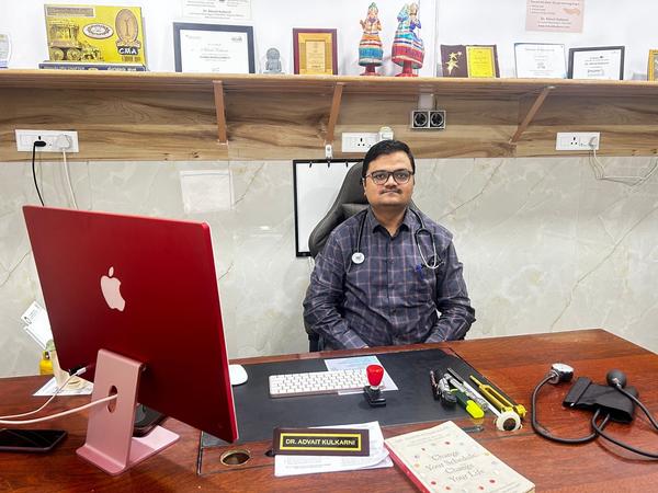

Dr. Advait Kulkarni (Neurologist)
neurologist near me
neurologist in bengaluru #neurologistinbengaluru
#neurologistnearme
Diagnosis
• A physical exam- Neurologist will do a number of tests you're familiar with, such as listening to the heart and checking the blood pressure.
• Blood tests- You may have several blood tests, including tests to check how fast the blood clots, whether the blood sugar is too high or low, and whether you have an infection.
• Computerized tomography (CT) scan- A CT scan can show bleeding in the brain, an ischemic stroke, a tumor or other conditions.
• Magnetic resonance imaging (MRI)- An MRI can detect brain tissue damaged by an ischemic stroke and brain haemorrhages.
• Carotid ultrasound- This test shows build-up of fatty deposits (plaques) and blood flow in the carotid arteries.
• Cerebral angiogram- In this uncommonly used test, your doctor inserts a thin, flexible tube (catheter) through a small incision, usually in the groin, and guides it through the major arteries and into the carotid or vertebral artery. Then your doctor injects a dye into the blood vessels to make them visible under X-ray imaging. This procedure gives a detailed view of arteries in the brain and neck.
• Echocardiogram- An echocardiogram can find a source of clots in the heart that may have travelled from the heart to the brain and caused a stroke.
neurologist near me
neurologist in bengaluru #neurologistinbengaluru
#neurologistnearme
Diagnosis
• A physical exam- Neurologist will do a number of tests you're familiar with, such as listening to the heart and checking the blood pressure.
• Blood tests- You may have several blood tests, including tests to check how fast the blood clots, whether the blood sugar is too high or low, and whether you have an infection.
• Computerized tomography (CT) scan- A CT scan can show bleeding in the brain, an ischemic stroke, a tumor or other conditions.
• Magnetic resonance imaging (MRI)- An MRI can detect brain tissue damaged by an ischemic stroke and brain haemorrhages.
• Carotid ultrasound- This test shows build-up of fatty deposits (plaques) and blood flow in the carotid arteries.
• Cerebral angiogram- In this uncommonly used test, your doctor inserts a thin, flexible tube (catheter) through a small incision, usually in the groin, and guides it through the major arteries and into the carotid or vertebral artery. Then your doctor injects a dye into the blood vessels to make them visible under X-ray imaging. This procedure gives a detailed view of arteries in the brain and neck.
• Echocardiogram- An echocardiogram can find a source of clots in the heart that may have travelled from the heart to the brain and caused a stroke.
Keywords